
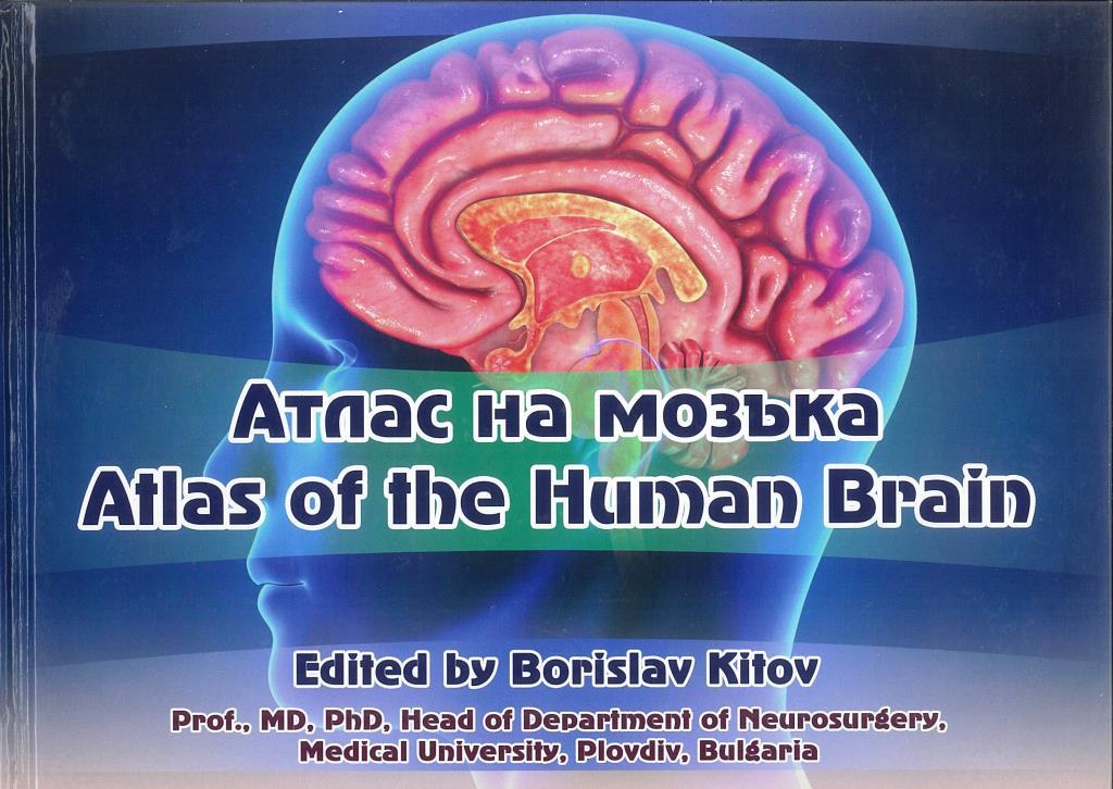
Atlas of the Brain
The atlas has been developed to help medical students, dentists and practitioners familiarize themselves with the capabilities of modern brain imaging so that they can orientate themselves in terms of the exact localization of the lesion and the type of structures involved, which will be helpful for them to make an accurate diagnosis.
- 9786191890286
- Atlas of the Brain
Netter Atlas of Human…
The atlas presents the anatomy of the brain not only in the traditional way, but also through computed tomography (CT) and magnetic resonance imaging (MRI), which have the unique visualization capabilities of both the respective intracranial lesions, as well as the affected substrate.
The atlas begins with the general anatomy of the brain, and subsequent axial, sagittal and coronal sections reveal in much greater detail its structure.