
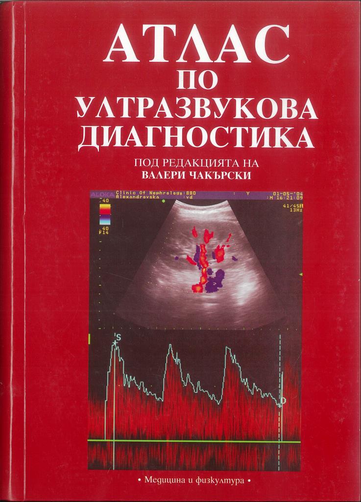
Atlas of Ultrasound Diagnostics
Preface
1. Ultrasound diagnostic equipment
2. Basic concepts and images in ultrasound. Principles of Doppler diagnostics. Contrast agents
3. Topographic anatomy
4. Liver
5. Doppler examination of the liver vascular system
6. Biliary system
7. Pancreas
8. Slezka
9. Kidneys
10. Doppler examination of the kidneys
11. Adrenal glands
12. Pathological processes outside the abdominal organs
13. Bladder
14. The prostate
15. Scrotum
16. The ultrasound method in pediatrics
17. Thyroid gland
18. Salivary glands
19. Breast gland
20. Thorax
21. Eye and orbit
22. Diagnostic Correlative Tables
Index
Summary
Summary
Multifunctional an atlas including both established techniques and novelties in the echographic diagnosis of diseases of the thorax, abdomen, the genitals, thyroid, thoracic and salivary glands, the eye; a chapter on ultrasound in pediatrics is also included.
=-=
Conventional ultrasound of the kidney is used commonly to depict structural abnormalities. It is limited, however, by a lack of functional and vascular information. Doppler sonography can reduce this limitation of standard sonography quickly and noninvasively.
Doppler examinations, although not difficult, must be done properly to obtain useful data. Information regarding the presence and direction of flow in renal vessels can be obtained. Vascular stenosis can be identified by several Doppler criteria, although the role of Doppler as a screening measure remains controversial. Assessment of vascular resistance is possible from Doppler waveform analysis, using parameters such as the resistive index. These data may provide hemodynamic and predictive information regarding a dilated collecting system identified by conventional ultrasound.
Analysis of the resistive index may also provide helpful clinical information in nonobstructive renal disease. In certain clinical settings, such analysis provides diagnostic data not readily available with other clinical and laboratory assessment methods. Pharmacologically stimulated renal Doppler examinations may lead to even greater benefits in the future. This article reviews renal Doppler ultrasound, including the physiological basis for Doppler examination, the technical principles of renal Doppler sonography, and the clinical applications of Doppler findings.