
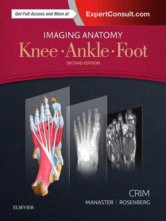
Imaging Anatomy: Knee Ankle Foot
Designed to help you quickly learn or review normal anatomy and confirm variants Imaging Anatomy: Knee Ankle Foot. Provides detailed anatomical views of each major joint of the lower extremity. Ultrasound and 3T MR images in each standard plane of imaging (axial coronal and sagittal) accompany highly accurate. Detailed medical illustrations assisting you in making an accurate diagnosis. Comprehensive coverage of the knee ankle and foot combined with an orderly easy-to-follow structure. It makes this unique title unmatched in its field.
Key Features
-
- Includes all relevant imaging modalities, 3D reconstructions. Highly accurate and detailed medical graphics that illustrate the fine points of the imaging anatomy
-
- Depicts common anatomical variants (both osseous and soft tissue) and covers imaging pitfalls as a part of its comprehensive coverage
-
- Enables any structure in the lower extremity to easily be located, identified, and tracked in any plane for a faster, more accurate diagnosis
-
- Provides richly labeled images with associated commentary as well as scout images to assist in localization
-
- Explains uniquely difficult functional or anatomical regions of the lower extremity. Such as posterolateral corner of knee, ankle ligaments, ankle tendons, and nerves of the lower extremity
-
- Presents coronal and axial planes as both the right and left legs, on facing pages, making ultrasound/MR correlation even easier
-
- Expert Consult™ eBook version included with purchase. This enhanced eBook experience allows you to search all of the text, figures, videos, and references from the book on a variety of devices.
- Imaging Anatomy: Knee Ankle Foot
Author Information
By Julia R. Crim, MD, Chief of Musculoskeletal Radiology Vice Chair for Clinical Affairs Professor of Radiology University of Missouri at Columbia Columbia, Missouri; BJ Manaster, MD, PhD, FACR, Emeritus Professor Department of Radiology University of Utah School of Medicine Salt Lake City, Utah and Zehava Sadka Rosenberg, MD, Professor of Radiology and Orthopedic Surgery NYU School of Medicine NYU Langone Medical Center New York, New York