
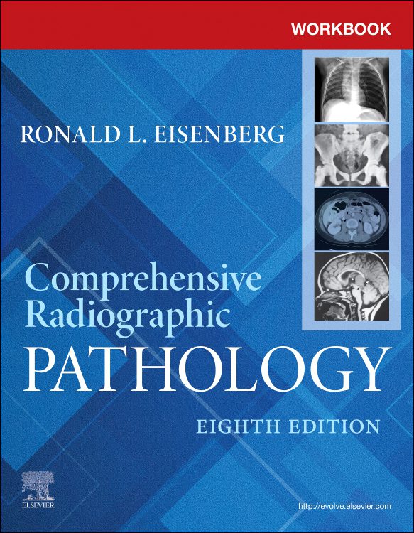
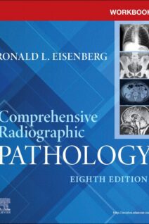
Workbook for Comprehensive Radiographic Pathology, 8th Edition
- NEW! Exercises on AI (artificial intelligence) and personalized medicine are added to this edition.
- NEW! Content on COVID as pertaining to chest X-rays is added.
- NEW! Updated questions are included in each chapter, as well as updated illustrations.
- Thorough review reflects the content in the Comprehensive Radiographic Pathology textbook and helps you understand disease processes, their radiographic appearance, and how to produce optimal diagnostic images.
- Wide variety of exercises includes fill-in-the-blank, matching, labeling, and multiple-choice questions, all designed to help you learn anatomy, identify pathology, and make technique adjustments.
- Case studies with diagnostic images make it easier to notice relevant details on the image and become familiar with the appearance of pathologies in different imaging modalities.
- Anatomic images let you review A&P and gain practice with labeling and analysis.
- Self-tests in each chapter include 20-40 multiple-choice questions, allowing you to assess your understanding of the material.
The Workbook for Comprehensive Radiographic Pathology is a comprehensive and practical guide to understanding and interpreting various radiographic techniques used in diagnostic pathology. The authors, Dr. Jeffrey A. Wright and Dr. James R. Armstrong, provide a systematic review of radiographic techniques, including X-rays, CT (CT), magnetic resonance imaging (MRI) and ultrasound, focusing on understanding their working principles, indications and limitations.
The textbook begins with an introduction to radiographic pathology, which provides an overview of the study of radiographic techniques in diagnostic pathology. It then moves on to the basic principles of each radiographic method, including a discussion of their physical properties, imaging techniques, and interpretation of the resulting images.
Subsequent sections of the textbook focus on specific applications of radiographic techniques in diagnostic pathology, discussing relevant indications, contraindications, and potential complications. In each chapter, the authors provide detailed information on the use of different radiographic techniques in different clinical scenarios, focusing on understanding their diagnostic capabilities and limitations.
One of the notable qualities of the Workbook for Comprehensive Radiographic Pathology is its emphasis on the practical application of radiographic techniques in diagnostic pathology. The authors integrate knowledge from various fields, including imaging, internal medicine, and surgery, to provide a comprehensive review of radiographic pathology.
Additional features included in this textbook further enhance its usefulness. These features include:
- Case studies: The textbook presents numerous real cases from clinical practice that illustrate the practical application of radiographic techniques in diagnostic pathology. These cases serve as valuable tools to stimulate critical thinking and encourage discussions about the choice of imaging technique and the interpretation of the resulting images.
- Online resources: Along with the print edition, an online study kit is available, which includes interactive exercises, self-assessment tests and additional information on selected topics. These digital resources complement the book's content and provide clinicians with a convenient way to review and apply their knowledge.
- Glossary: The textbook includes a comprehensive glossary that defines key terms used throughout the text. This reference helps clinicians quickly look up and understand unfamiliar terms while reading.
Overall, the Workbook for Comprehensive Radiographic Pathology is a comprehensive and well-organized guide to understanding and interpreting various radiographic techniques used in diagnostic pathology. Its combination of solid scientific material, clinical applications and diverse features make it an indispensable tool for both clinicians involved in diagnostic pathology and students and trainees who wish to improve their understanding of radiographic pathology and develop selection and interpretation skills of radiographic techniques. Whether used as a primary textbook or a reference, this textbook will undoubtedly contribute to a deeper understanding of the world of radiographic pathology and its applications in modern medicine.